Imaging and Engineering Core
Core Leadership and Personnel
The Co-Directors of the Imaging and Engineering Core are Drs. Mihaela Balu and Scott Atwood. Dr. Mihaela Balu, PhD, directs the imaging core and is the leader of the Nonlinear Optical Microscopy (NLOM) Lab, an imaging facility highly focused on applications related to in vivo and ex vivo skin imaging in both humans and animal models. Dr. Scott Atwood, PhD, directs the skin organoid engineering core and is an expert in skin biology and skin organoids.
Contacts
Users are encouraged to make an appointment with Amanda Durkin (afdurkin@uci.edu) for using the imaging resources available at BLI and with Scott Atwood (satwood@uci.edu ) for skin organoid engineering resources.
Additional information about submitting orders is available on the NLOM website and through iLab.
Skin Organoid Technologies
The Engineering Core uses a classic human skin equivalent (HSE) organoid model. Contemporary skin models predominantly comprise two discernible layers: the epidermis and dermis. This design allows for the optimal differentiation of the epidermis and the replication of the complex interactions between keratinocytes and fibroblasts, which are crucial for maintaining skin homeostasis. These reconstructed skin models are composed of an extracellular matrix-based biomaterial, such as collagen or devitalized dermis, which is primarily populated by fibroblasts and overlaid with a stratified epidermis. This structural arrangement ensures that the dermal layer remains in direct contact with the culture medium, while the epidermis is exposed to the air. Collagen- and devitalized dermis-based HSE organoids are increasingly recognized as promising skin models in skin bioengineering owing to their supportive cellular environments and low antigenicity. HSEs are used to investigate various aspects of normal and abnormal skin biology, including wound healing, aging, and various skin diseases. Additionally, they have been employed directly in studies and as "hybrid" models, where humanized HSEs are grafted onto immunodeficient mice. Furthermore, in response to challenges associated with donor variability, conventional primary cell-based HSEs have transitioned to more standardized and reproducible in vitro culture models. These models utilize either immortalized cell lines or cells derived from hiPSCs.
Imaging Technologies
The Imaging Core includes the following technologies at BLI:

Leica SP8/FILM/CARS
Leica SP8/FLIM/CARS. Leica SP8 is customized to feature the following imaging modalities: confocal fluorescence and two-photon excited fluorescence (TPEF) microscopy, second and third harmonic generation (SHG/THG), coherent anti-Stokes Raman Scattering (CARS) and fluorescence lifetime microscopy (FLIM). Available excitation light sources: Insight X3 laser (SpectraPhysics) with dual output: 680-1300 nm, 120 fs and 1045 nm, 170 fs; cw lines and a white-light laser source for confocal fluorescence microscopy. Suitable for imaging of cells, tissue and animal models (in vivo).
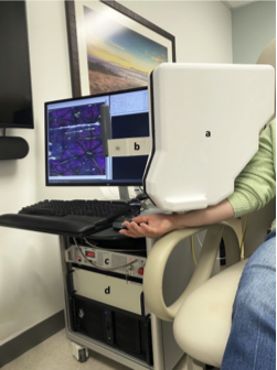
In vivo imaging of human skin using FLAME
Fast Large Area Multiphoton Exoscope (FLAME). FLAME is a portable, custom developed multiphoton microscopy platform, highly optimized for efficient in vivo imaging of human skin in clinical setting. It provides contrast based on intensity and time-resolved label-free two photon excited fluorescence from keratin, NADH/FAD, melanin and elastin fibers. Collagen fibers are visualized through second harmonic generation detection. The studies on this system are performed based on approved IRB protocols.
Spatial Frequency Domain Imaging (SFDI). The available device based on the SFDI technology is a commercial imaging platform (Modulim, Irvine, CA) that enables quantitative, longitudinal assessment of cutaneous hemodynamics to diagnose disease and monitor therapy up to 5 mm in depth beneath the surface of skin. This device is available in Prof. Anthony Durkin’s SFDI Lab at BLI.
Laser Speckle Imaging (LSI). The available device based on the LSI technology is a handheld instrument that measures skin blood flow at depths of ~0.5 mm. This device is available in Prof. Bernard Choi’s LSI Lab at BLI.
Click here for a complete list of the BLI shared resources and details on requesting service.
Workshops, Tech Talks, and Seminars
The FLuorescence Advanced Imaging Research (FLAIR) Workshop at UC Irvine brings together cutting-edge developments in optical fluorescence microscopy, metabolic imaging, probe design, and AI-driven image analysis. FLAIR merges two long-standing microscopy workshops, the UCI Laboratory for Fluorescence Dynamics (LFD) and UC Berkeley's Advanced Imaging Methods Workshop (AIM), into a single annual large event, fostering collaboration and innovation. More information can be found here link.
The Beckman Laser Institute, through its LAMP seminar series, offers weekly talks on the latest imaging methods. Information about past and upcoming seminar can be found here. Please, contact Erin Miller (ewmiller@uci.edu) at BLI if you wish to be added to the mailing list for learning about these seminars.